
When Will 3D Printed Organs Become a Reality?
A process engineer's read on 3D printed organs: which methods exist, where they break, and what is already implantable. Written for engineers and buyers who need to separate lab results from production reality.
In this article
- 1
- 2
- 3
- 4
- 5
- 6
- 7
Key takeaways
How 3D printed organs are actually built
Bioprinting borrows the motion platform of a CNC machine and swaps the cutter for a deposition head. A gantry or robotic arm moves in X, Y, and Z while a nozzle, inkjet head, or light projector places material. The material is not plastic. It is a hydrogel loaded with living cells, usually suspended in a support bath so the soft structure does not collapse under its own weight.
The build starts from imaging. CT or MRI data is segmented into a digital model, then sliced into toolpaths. Slice height for extrusion bioprinting typically runs 100–300 μm, close to what a fused-deposition printer uses. The difference is the payload: each deposited line carries cells at 1–10 million cells per mL, and those cells must survive the shear stress of passing through a 200–500 μm nozzle.
After printing, the construct goes into a bioreactor. Perfusion, temperature at 37 °C, and oxygen tension are controlled for days to weeks. This maturation step is where most failures happen. A construct that looks perfect on the print bed can delaminate, calcify, or lose cell viability once it is perfused at physiological flow rates.
So the honest answer to when will 3D printed organs become a reality is not one date. It is a ladder. Flat tissue is on the ladder's first rung. Hollow tubes are on the second. Solid organs with a full capillary bed are still several rungs up.
- 1Motion platformGantry, delta, or 6-axis arm; positional accuracy matters more than speed.
- 2Bio-inkHydrogel plus cells; viscosity and crosslink speed set printability.
- 3Support bathKeeps overhangs and thin walls from slumping before crosslinking.
- 4BioreactorPerfusion and conditioning decide whether the tissue matures or dies.
Three printing methods and where each one fits
Extrusion bioprinting pushes bio-ink through a nozzle with pneumatic or screw pressure. It handles high cell densities and viscous hydrogels, and it is the cheapest route to a centimeter-scale construct. The trade-off is resolution: lines land at 200–500 μm, and cells near the nozzle wall see the highest shear. Viability after printing often sits between 80% and 95% depending on nozzle diameter and pressure.
Inkjet and laser-assisted methods drop or transfer picoliter volumes. Resolution improves to roughly 10–50 μm, which is close to a single cell. Throughput drops sharply, and the inks must be low-viscosity, so you cannot print a dense collagen matrix this way. These methods suit thin sheets and patterned co-cultures, not a whole kidney.
Light-based methods such as digital light processing cure a photosensitive hydrogel layer by layer. A full layer appears in one exposure, so speed scales with height rather than volume. Vat-based systems have produced small vascular networks and heart-valve geometries. The chemistry is the limit: photoinitiators and UV exposure can damage DNA, so cell survival depends on wavelength, dose, and photoinitiator choice.
None of these methods is a general answer. Pick extrusion for volume and cell density. Pick jetting for fine patterning. Pick light-based printing when you need a dense capillary tree inside a soft matrix. Most serious research programs run two of the three.
- 1Extrusion200–500 μm lines; high cell density; best for centimeter-scale tissue.
- 2Inkjet / laser10–50 μm features; low-viscosity ink only; best for thin patterns.
- 3Light-basedOne exposure per layer; fast for tall parts; UV dose limits cell survival.
Why vascularization blocks every solid 3D printed organ
Every living cell sits within 100–200 μm of a capillary. That is the diffusion limit for oxygen. A solid organ is centimeters thick, so a printed organ needs a branching vessel tree that reaches every cell, from a 5 mm artery down to a 5 μm capillary. Printing that tree is the hard part, and keeping it open after printing is harder.
The engineering problem has three layers. First, resolution: extrusion cannot draw a 5 μm channel. Second, mechanics: a soft hydrogel channel collapses under 100 mmHg of arterial pressure unless the wall is reinforced. Third, biology: endothelial cells must line the channel and stay attached under shear. Miss any one layer and the core of the construct dies within hours.
Research groups attack this from different angles. Some print a sacrificial ink, cast the hydrogel around it, then dissolve the ink to leave channels. Others print a rigid scaffold with machined or molded channels and seed cells into it. A third route bioprints directly onto a perfusable chip and keeps the tissue alive in vitro rather than implanting it.
That third route matters commercially. A liver model on a chip that stays viable for 30 days has real value for drug testing, and it does not need an implant approval. It is a stepping stone, not a compromise.
- 1Diffusion limitOxygen reaches roughly 100–200 μm from a capillary before cells starve.
- 2Pressure limitUnreinforced hydrogel channels collapse at arterial pressures.
- 3Shear limitEndothelial lining must survive flow without detaching.
What is already implanted in patients
Skin is the clearest success. Bioprinted skin substitutes with a printed dermal layer and a keratinocyte top layer have been used in burn and chronic-wound care. The construct is flat and thin, so the vascular problem is smaller: the wound bed supplies the blood. A 2022 case in San Antonio, Texas, implanted a custom external ear in a 20-year-old woman born without ears, matched to the size and shape of her other ear.
Bladder patches and cartilage templates follow the same logic. They are avascular or nearly so, and they rely on surrounding tissue for nutrients while they integrate. Ear and nose cartilage is a good fit because the shape is the function. If the printed geometry matches the patient's anatomy, the mechanical requirement is met.
Bone is advancing on a different track. Printed calcium phosphate and titanium scaffolds act as a frame that the body fills in. Here the printer makes the scaffold, not the living tissue. That distinction keeps the regulatory path simpler and the load-bearing requirement manageable.
So the pattern is clear. Thin, avascular, or non-load-bearing structures are implantable now. Thick, vascular, contractile organs are not. A printed ear is reality. A printed kidney is a research program.
- 1SkinPrinted dermal and epidermal layers used in wound care.
- 2CartilageEar and nose shapes where geometry equals function.
- 3Bone scaffoldPrinted frame that the body remodels into bone.
- 4Not yetKidney, liver, heart, and lung remain preclinical.
Bio-inks, scaffolds, and the sterilisation step
A bio-ink is judged on four things: printability, crosslink speed, mechanical stiffness after curing, and cytocompatibility. Alginate crosslinks fast with calcium and prints cleanly, but mammalian cells do not attach to it well. Collagen and fibrin give cells real attachment sites, but they are soft and slow to gel. Gelatin methacryloyl sits in the middle and cures under light in seconds. Most published work blends two or three of these.
Stiffness matters more than people expect. Cells read the stiffness of their surroundings and change behavior accordingly. A hydrogel at 0.5–3 kPa behaves like soft tissue; above 20 kPa it starts to look like scar. If the printed matrix is too stiff, the cells do not form the tissue you intended, even if the geometry is perfect.
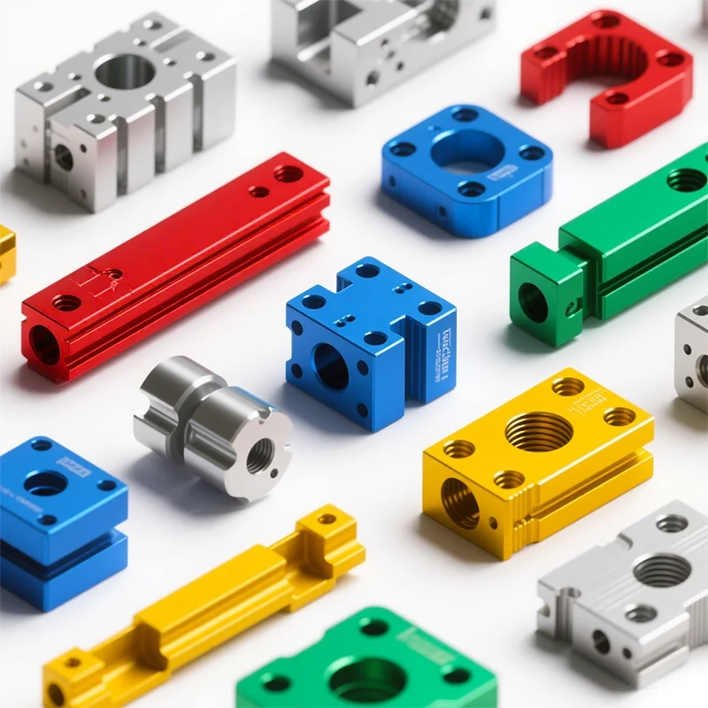
The scaffold is often not printed at all. It is a machined or molded frame made from medical-grade polymer, titanium, or stainless. For a research fixture or a perfusion housing, that frame is a machined part with tight tolerances, and it must survive autoclave cycles at 121 °C or gamma sterilisation without warping.
Sterilisation is a hard filter on material choice. Autoclave heat degrades many hydrogels. Gamma radiation can crosslink or chain-scission a polymer. Ethylene oxide needs aeration time to clear residue. The material and the sterilisation method have to be chosen together, not one after the other.
- 1AlginateFast calcium crosslink; prints well; weak cell attachment.
- 2Collagen / fibrinNative attachment sites; soft; slow gelation.
- 3GelMALight-cured in seconds; stiffness tunable by degree of substitution.
- 4Stiffness window0.5–3 kPa mimics soft tissue; above 20 kPa reads as scar.
Which bioprinting method fits which target
Feature size and cell density decide the method. No single platform covers every organ.
| Method | Feature size | Cell density | Best fit |
|---|---|---|---|
| Extrusion | 200–500 μm | High, 1–10 M cells/mL | Centimeter-scale tissue, skin, cartilage |
| Inkjet / laser | 10–50 μm | Low to medium | Thin sheets, co-culture patterns |
| Light-based (DLP) | 10–50 μm | Medium | Dense capillary trees, valve geometry |
| Sacrificial ink + cast | 50–200 μm channels | Set by casting resin | Perfusable vessel networks |
| Machined scaffold + seeding | 100 μm and up | Seeded after build | Bone, load-bearing frames, fixtures |
What to expect and when
If you need flat, avascular, or non-load-bearing tissue, 3D printed organs are already a reality and shipping. If you need a solid vascular organ like a kidney or heart, plan for a decade of preclinical work before the first routine implant. Choose the chip-and-scaffold route for drug testing today, and treat whole-organ printing as a research bet.
Questions engineers keep asking
Is any 3D printed organ approved for routine transplant?
No solid organ is approved for routine transplant. Skin substitutes, cartilage templates, and bladder patches have reached clinical use, and a custom external ear was implanted in 2022 in San Antonio, Texas.
Those cases work because the tissue is thin, avascular, or non-load-bearing. Solid organs still fail on vascularization.
Why is a printed kidney harder than a printed ear?
A kidney is centimeters thick and filters blood at arterial pressure. Every cell needs a capillary within 100–200 μm, and the vessel walls must hold pressure without collapsing.
An ear is avascular and carries no flow. Geometry is the main requirement, so the biological load is far lower.
What tolerances and surface finishes matter on the hardware side?
Perfusion housings and scaffold frames usually need ±0.005 mm on mating features and Ra 0.8–1.6 μm on wetted surfaces. Rough surfaces trap cells and complicate cleaning.
We machine these frames from titanium, stainless 316L, or medical-grade polymer, and inspect 100% before shipment.
Can a 3D printed organ model replace animal testing?
Partly. A liver or kidney model on a perfusable chip can stay viable for weeks and is useful for toxicity screening.
It cannot yet reproduce whole-organ pharmacokinetics. Most groups treat it as a complement to animal studies, not a replacement.
Which sterilisation method survives a printed construct?
It depends on the matrix. Autoclave at 121 °C destroys most hydrogels. Gamma radiation can chain-scission polymers. Ethylene oxide needs aeration to clear residue.
Decide the sterilisation route before you pick the material, or you will reprint the whole batch.
How does GreatLight fit into bioprinting work?
We machine the rigid parts: perfusion housings, scaffold frames, bioreactor fixtures, and test rigs. We hold ±0.005 mm and work from one prototype to 10,000+ parts with no minimum order quantity.
We also run ISO 13485:2016 for medical device work and sign NDAs on request. Uploads stay confidential.
Need machined hardware for your bioprinting rig?
Send your drawings and we will return a quote with a free DFM analysis within 12 hours. Prototypes and production runs use the same inspection route.
12-hour quote100% inspectionNo minimum orderISO 13485:2016
More process notes
We publish setup notes, tooling trials and inspection data from the factory floor.











