
Breakthrough: The First 3D Printed Cornea Tested on Animals
In 2022 a team at LV Prasad Eye Institute in India printed a cornea from bioink and transplanted it into rabbit eyes. This page covers what was actually printed, why the gel formulation matters more than the printer, and how the same process limits show up in medical hardware you would machine instead of print. Written for engineers and sourcing staff who need to decide where additive stops and subtractive starts.
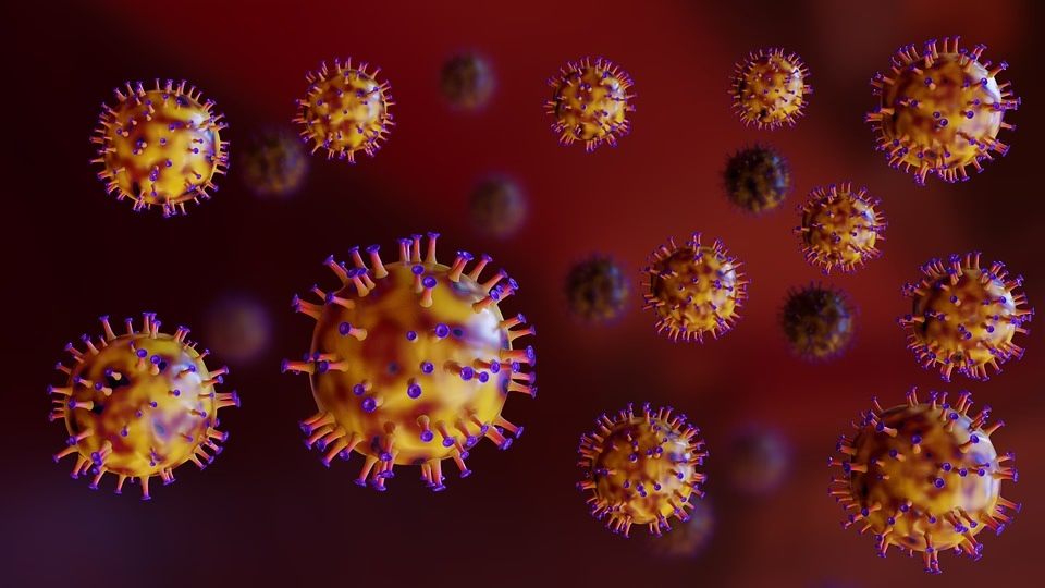
A printed cornea and the tooling questions it raises
Start with what the 2022 trial proved, then read the process limits that decide whether a part is printed or machined.
The first 3D printed cornea: what the team actually transplanted
On 17 August 2022, a research group at the LV Prasad Eye Institute in Hyderabad, working with the Indian Institute of Technology, the University of Hyderabad and the Centre for Cellular and Molecular Biology, reported that a 3D printed cornea had been successfully tested on animals. The construct was transplanted into rabbit eyes. Rabbits are a standard early model for corneal work because their eyes are close enough in curvature and layer thickness to human tissue for a short-term tolerance check.
The printed part was not a clear plastic lens. It was a hydrogel disc built layer by layer from a bioink that carries live cells. The team described the material as free of residual donor cornea tissue, which is the point of the work: a printed cornea does not depend on a cadaver donor supply.
The printer itself is the least interesting component. A multi-nozzle extrusion head deposits a viscous gel through a fine tip, usually in the 200 to 600 μm range, and the gel must hold its shape after the tip passes. That is a rheology problem, not a motion-control problem.
Why the bioink decides the outcome, not the machine
A cornea is roughly 0.5 mm thick at the center and about 11.5 mm across. Five layers run from epithelium to endothelium, and the stroma in the middle makes up most of the thickness. Printing that stack means holding dimensional accuracy while the gel is still wet, then keeping the cells alive through the whole build.
Cell viability drops with time under the nozzle and with shear stress in the syringe. A print that takes 40 minutes may lose far more cells than one that takes 15 minutes, even with identical geometry. Formulation chemists tune viscosity, crosslinker concentration and curing wavelength to shorten that window.
Optical clarity is the harder target. Light has to pass through the construct without scattering, so any entrapped bubble, unmixed blob or layer seam shows up as haze. In a machined optical part we would talk about Ra 0.2–0.8 μm and surface figure; in a printed hydrogel the equivalent problem is refractive index uniformity across the disc.
- 1Extrusion tipTypically 200–600 μm; smaller tips raise shear stress and cut cell survival.
- 2Build timeMinutes matter. Long builds cost viability even when geometry is fine.
- 3CrosslinkingUV or chemical cure must set the gel without damaging the embedded cells.
- 4SterilityThe whole path, ink to nozzle to dish, has to stay clean.
When a printed part makes sense and when it does not
Additive wins when the geometry is organic, internal channels are needed, or the part count is one. A patient-specific implant that matches a scan is a natural print. So is a lattice that would be impossible to cut with a tool.
Subtractive wins when the material is metal, the tolerance is tight, or the surface has to be optical or sealing. A titanium fixation plate, a stainless instrument body or a machined lens mold insert will not come off a polymer printer. Those parts need 5-axis work, and the finish has to be measured, not eyeballed.
The practical rule is simple. If the part carries load, seals a fluid, or has to hold ±0.005 mm, machine it. If it is a soft structure with a biological interface and low stiffness, print it. When a device needs both, split it into a printed soft component and a machined rigid frame, then join them in a clean process.
Printed hydrogel versus machined hardware
Two process families that share a device but not a tolerance budget.
| Attribute | 3D printed hydrogel | Machined metal or plastic |
|---|---|---|
| Typical material | Cell-laden bioink | Aluminium, stainless, PEEK, PMMA |
| Wall or feature size | 200–600 μm nozzle limited | Down to 0.5 mm with the right tool |
| Achievable tolerance | Printer and shrink dependent | ±0.005 mm |
| Surface finish | Haze from layer seams | Ra 0.2–0.8 μm when specified |
| Best use | Soft, patient-specific structures | Load, seal and optical parts |
| Sterilization | Gentle, low temperature | Autoclave, gamma or EtO |
What the trial means for a medical device program
A rabbit trial is a tolerability result, not a product. It tells you the printed gel did not trigger an obvious rejection over the study window. Human use needs much longer data, a repeatable print recipe, and a release test that proves every disc is the same.
That release test is where machining experience helps. A printed cornea still needs a mold or a fixture, a carrier for handling, and a sterile package. Those parts are dimensioned and inspected like any other hardware. A transparent carrier with a defined curvature is a CNC job, and it is usually the first thing to get wrong.
We run ISO 13485:2016 alongside ISO 9001:2015, IATF 16949:2016 and ISO 27001:2022, and we inspect 100% of parts before shipment. If your program needs machined tooling, fixtures or implant-grade components around a printed structure, that is the work we quote.
Send a drawing or a STEP file. Uploads stay confidential, and an NDA is available on request.
Questions engineers ask next
Was the 3D printed cornea tested on humans?
No. The 2022 work reported by the LV Prasad Eye Institute team was an animal trial in rabbit eyes. It was a short-term tolerability and tissue-response check.
Human trials need longer follow-up, a validated print recipe and a release specification for every construct.
What material was used?
The construct was built from a bioink, a cell-carrying hydrogel, and the team stated it contained no residual donor cornea tissue. It is not a clear polymer or a machined lens.
Hydrogel chemistry varies by group: collagen, alginate, gelatin derivatives and blends are all used.
Can a cornea be CNC machined instead?
Not a live one. Machining removes material and kills cells at the cut. But the mold inserts, carriers and fixtures used to handle and shape printed tissue are machined.
Those parts can be held to ±0.005 mm and finished to Ra 0.2–0.8 μm when the optical surface matters.
Which medical hardware do you actually machine?
Instrument bodies, fixation plates, housings, manifolds, mold inserts, test fixtures and carriers. Materials include 316L, 17-4PH, titanium Ti-6Al-4V, PEEK and PMMA.
We hold ±0.005 mm and inspect 100% of parts before shipment, with reports on request.
How fast can a medical prototype be quoted?
Quotation and a free DFM analysis come back within 12 hours. Production can start within 24 hours of approval, and parts ship in 3–5 days.
There is no minimum order quantity, so a single prototype is fine.
How is confidentiality handled?
Uploads are secure and confidential. We sign an NDA on request before drawings change hands.
We hold ISO 27001:2022 for information security management.
Need the machined parts around a printed structure?
Send a drawing or STEP file and we will return a quote with a free DFM analysis within 12 hours.
12-hour quote100% inspectionNDA on request









